More Information
Craniosynostosis
What is craniosynostosis?
Craniosynostosis (CRA-nee-oh-sin-us-TOE-sis) refers to the premature closure of the growth centers between bones in a baby’s head. It happens too early in the baby’s development, before the baby’s brain has finished growing.
The human skull is made up of five bones. In newborns, these bones are not fully joined into a solid surface. Flexible connective tissue called sutures hold the five bones together along their edges. Also, normally a baby has two soft spots, called fontanelles, on the head. The anterior fontanelle is near the front of a baby’s head on top; the posterior fontanelle is on the top closer to the back. These soft spots are spaces where two or more of the bones of the skull meet.
As a baby grows, so does the brain. The soft spots and flexible sutures give the baby’s brain room to grow. The skull does not complete growth and the sutures remain open until teenage years, although the soft spots often close in the first several years of life.
When craniosynostosis occurs, it prevents the skull from growing in the area where the early bone closure is located. This can result in the baby’s skull growing into an abnormal shape. It can also put pressure on the brain, a condition called intracranial pressure. The consequences of this are not well understood, but may be associated with issues later in life.
With craniosynostosis, a baby’s brain will grow in the direction of places where the bones of the skull have not yet fused and there is flexibility. In simple terms, If the brain can’t grow upward because bone prevents it, it will expand outward.
Ask your pediatrician about head shape concerns
It is not unusual for a baby’s head to be a little pointy or cone-shaped after a vaginal delivery. In a few days to a couple of weeks, it will usually round out on its own.
But if you have any concerns about how your baby’s head looks, talk with your pediatrician. Your baby’s doctor will likely check the head’s shape during routine checkups. If a baby does have craniosynostosis, most experts believe that treatment should begin during the first year of life. Early treatment usually allows for a better outcome. It may also reduce the risk of developing issues later in life.
If you want to discuss your concerns with a pediatric craniofacial specialist at Children’s of Mississippi, you can schedule an appointment online. Schedule an appointment.
What causes craniosynostosis
Scientists are not sure what causes craniosynostosis, although it is likely related to more than one thing. Some evidence suggests genetics plays a role. Some types, called syndromic craniosynostosis, occur with abnormalities in other parts of the body as well. The most common syndromes that include craniosynostosis include Apert, Crouzon, Pfeiffer, Muenke, and Saethre-Chotzen syndromes.
How it is diagnosed
In most cases, craniosynostosis is diagnosed soon after a baby is born, usually with a physical examination. An unusual shape to the baby’s head is the most common symptom. Your doctor may examine your baby’s skull to find out if the “soft spot” is missing or unusual. There may be hard spots in one or more sutures causing a raised ridge. Further testing, such as CT scans, may be needed to confirm the diagnosis and discover how much bone has fused and in what specific areas.
Types of craniosynostosis
There are several types of craniosynostosis, defined by which suture or sutures closed prematurely.
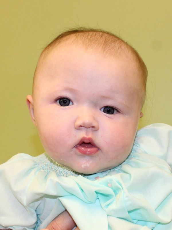
Sagittal craniosynostosis results from bones fusing at the sagittal suture, located on top of the head running from front to back. In this type, the baby’s head becomes long and narrow. This shape is called scaphocephaly. Sagittal craniosynostosis is the most common type.
Coronal or bicoronal craniosynostosis occurs when one or both of the coronal sutures close too early. There are two coronal sutures, right and left. They form a line from the top of the ear to the top of the head, connecting to the sagittal suture. Together, they make a plus-sign shape at the top of the head, with the anterior fontanelle (soft spot) in the center. When one of the coronal sutures closes too early, it results in a flat shape to the baby’s forehead on the side that fused. The eye socket could be raised and the nose could also be pulled to one side. Also called anterior plagiocephaly, this is the second most common type of craniosynostosis.
When both coronal sutures close too soon, called bicoronal craniosynostosis or brachycephaly, the baby’s head grows wide and short.
Metopic craniosynostosis results from bone fusing at the metopic suture, which runs upward from the baby’s nose to the sagittal suture on top of the head. This condition is also called trigonocephaly. When it occurs, the baby’s head may be narrow, almost pointed, in the front, and wide in the back.
Lambdoid craniosynostosis occurs at the lamboid suture, which runs along the back of the head. If this suture closes too early, the baby’s head may be flat on the back. The condition is also called posterior plagiocephaly. This type of craniosynostosis is extremely rare. It can look like a condition called deformational plagiocephaly.
Deformational plagiocephaly, sometimes called positional plagiocephaly or flat head syndrome, is not a type of craniosynostosis. This condition is very common. It can occur when a baby stays in the same position too long, always sleeping on the same side, for example. A craniofacial specialist can confirm this condition with a physical exam, and it can be treated without surgery. A baby may need to wear a special helmet until the head’s shape is corrected. Physical therapy may be needed if the condition is caused by tight muscles in the baby’s neck.
Treatment for craniosynostosis
In general, the treatment for craniosynostosis is surgery, with or without additional treatments. The goal of treatment is to correct head and facial differences and to relieve pressure on the baby’s growing brain, reducing the likelihood of developing issues later in life.
The type of surgery and treatment a baby needs depends on the type of craniosynostosis and its severity.
Broadly speaking, surgeries to treat craniosynostosis fall into two categories:
- Endoscopic minimally invasive procedures
- Open cranial vault remodeling (skull reshaping)
In general, minimally invasive procedures are done when a baby is between 3 and 6 months old. Open procedures are usually done when children are between 8 and 11 months old. The type of procedure your Children’s of Mississippi craniofacial team may recommend depends on several factors, all taking into consideration what will give your child the best possible outcome.
Common types of minimally invasive procedures include:
- Strip craniectomy (with or without a modeling helmet): In this procedure, the surgeon makes small incisions in the scalp and then uses an endoscope to remove a strip of bone from the skull, often the suture that has closed prematurely. An endoscope is a very thin flexible tube with a light and camera mounted on the end. The camera allows the surgeon to see into the space to perform the operation.
- Spring-mediated cranial vault remodeling: Often used in connection with a strip craniectomy, in this procedure the surgeon places spring-like devices onto the baby’s skull, expanding across the gap where the craniectomy was performed and connecting the bones. These springs allow the baby’s skull to expand in a normal direction and shape and give the baby’s brain room to grow. The springs are removed later in another surgery after the baby’s head has grown to the correct shape.
- Distraction-mediated cranial vault remodeling: In this procedure, a surgeon makes a cut into the skull and then places a metal device called a distractor into the bones to push them apart. Over time, the distractors are gradually widened, triggering the bones to grow to fill in the gap. This slow process for reshaping the skull into the desired shape uses the body’s ability to form bone, called osteogenesis, as part of the treatment.
In open procedures, the surgeon makes an incision into the scalp to access the part of the skull that needs reshaping. Then, the surgeon repositions the bones to achieve the correct shape. In most cases, the surgeon will overcorrect the shape to allow for head growth. The surgeon uses special plates and screws to hold the bones in the new shape. These are resorbable, meaning the body eventually absorbs them. This does away with the need for another surgery later to remove them.
There are many types of open surgery. One is called Fronto-Orbital Advancement (FOA).
It is used to treat bones of the forehead and eye sockets. The surgeon makes an incision from ear to ear across the top of the baby’s head. Then, the bones are shaped into the correct position and held together with resorbable plates and screws.
Procedures commonly offered at Children’s of Mississippi for craniosynostosis include:
Sagittal craniosynostosis:
- Strip craniectomy
- Endoscopic strip craniotomy with postoperative helmet therapy
- Strip craniectomy with placement of transutural springs
- Subtotal calvarial vault remodeling
- Fronto-orbital advancement
Coronal craniosynostosis:
- Strip craniectomy
- Endoscopic strip craniotomy with postoperative helmet therapy
- Strip craniectomy with placement of transutural springs
- Distraction osteogenesis
- Fronto-orbital advancement
Metopic craniosynostosis:
- Strip craniectomy
- Endoscopic strip craniotomy with postoperative helmet therapy
- Strip craniectomy with placement of transutural springs
- Fronto-orbital advancement
Lamboid craniosynostosis:
- Strip craniectomy
- Endoscopic strip craniotomy with postoperative helmet therapy
- Strip craniectomy with placement of transutural springs
- Posterior cranial vault remodeling
Can a baby with craniosynostosis live a normal life?
Every baby is unique and every case of craniosynostosis is different. If only one suture is involved, children most often develop normally and lead a normal life. Because it is difficult to measure intracranial pressure in babies and how that may affect brain growth, experts recommend keeping a watch on cognitive development. For this reason, patients with craniosynostosis are followed annually by the Children’s of Mississippi craniofacial team.
When more than one suture is involved or craniosynostosis is associated with a syndrome, there is a higher likelihood of raised intracranial pressure. These patients will be followed more closely by the team to make sure outcomes are the best possible.
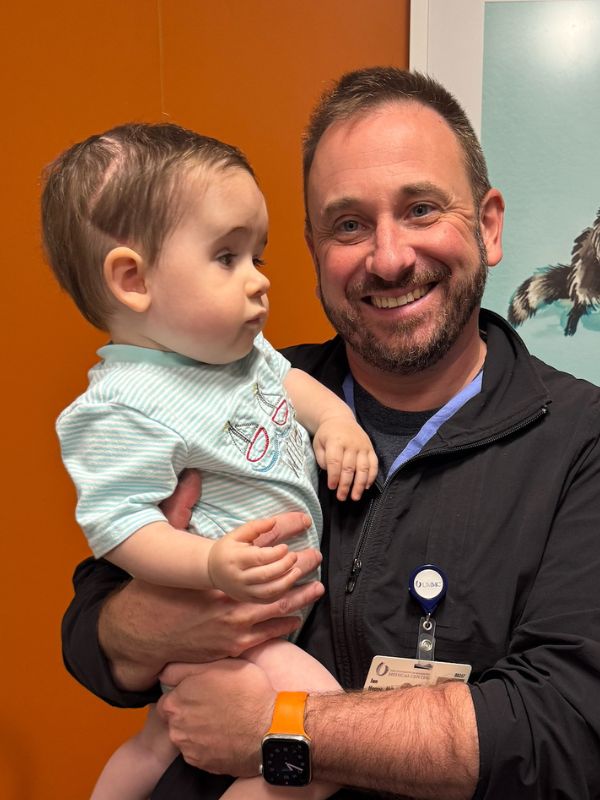
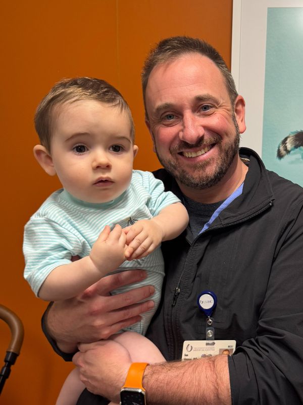
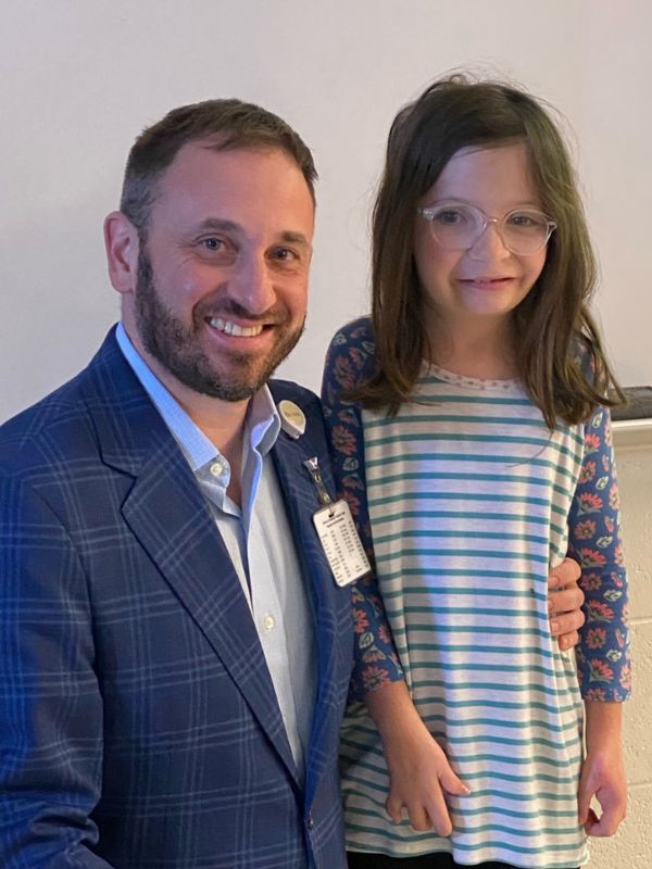
Children’s of Mississippi also hosts an annual reunion for kids and families who have had craniofacial surgery. It’s a great way for kids to make friends with others who share their experience, and for parents to share helpful tips and resources.
Get help at Children’s of Mississippi
If you are concerned about your child, you can request an appointment with a craniofacial specialist at Children’s of Mississippi. Request an appointment online.
Last reviewed 7/2/2025